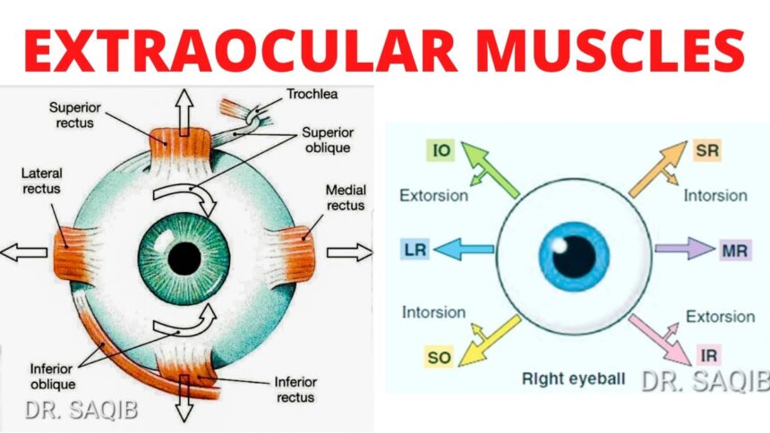
We’re now going to talk about clinically testing the extraocular muscles.
Extraocular muscles
This picture shows the anatomical actions on the left of the screen, the clinical testing of muscles on the right side of the screen. And what is the difference? That’s the purpose of this article. The medial rectus and lateral rectus muscles are the only two extra ocular muscles that act in the x axis.
So how will you clinically test these two muscles and their associated cranial nerves? To do that we take and look at their actions. Well the lateral rectus abducts the eye. The medial rectus abducts the eye. So to clinically test them have them look towards their nose or towards the wall. That’s simple.
Medial rectus will test cranial nerve three. Lateral rectus will test the abducens nerve. Cranial nerve six. The superior rectus and inferior oblique muscles are the only two extra ocular muscles that act in the y axis to elevate the eye and look up. Therefore, to test the superior rectus muscle, it must be isolated from the inferior oblique and vice versa. How is this done? Well, the superior rectus muscle is shown here in pink, and the inferior oblique is grayed out.
In order to isolate the superior rectus from the inferior oblique muscle, the vector pull of the muscles, the solid arrow, must be placed in parallel with the gaze of the orbit, the dotted arrow, and what action would be necessary in order to accomplish this? Correct. You have the patient abduct their eye. Now the vector pull of the superior rectus solid arrow is in parallel with the gaze of the orbit, the dotted arrow, and now the patient is instructed to look up.
Now what axis would the inferior oblique muscle now act if it contracted with the eye abducted? It’s on the z axis. It talks the eye. And that’s something that’s not easy to see. Now the inferior oblique muscle is highlighted with the superior rectus muscle grayed out. In order to isolate the inferior oblique from the superior rectus muscle, the vector pull of the muscle, that solid arrow must be placed in parallel with the gaze of the orbit. The dotted arrow. So what action would be necessary in order to accomplish this? Correct. They abduct their eye.
Also Read….
- Labetalol 100 mg (Trandate): Uses, Dosage and Side Effects
- Temazepam 15 mg (Restoril): Uses, Doge, Side Effects
- Fexofenadine 180 mg Uses Dose Side Effects
Now the vector pull of the inferior oblique. The solid arrow is in parallel with the gaze of the orbit. That dotted arrow. Now the patient is instructed to look up. So what axis would the superior rectus now act with the eye adducted it’s going to act on that z axis to talk the eye, which is not something easy to see. So in clinically testing, first you have the patient look laterally and then look up.
That’s how you isolate the superior rectus away from the inferior oblique. And to test the inferior oblique you have the patient adduct the eye first and then look up. And that’s the way you isolate the inferior oblique away from the superior rectus. So the superior oblique and the inferior rectus muscles are the only two extraocular muscles that act in the y axis to depress the eye and look down.
Therefore, to test the superior oblique, it must be isolated from the inferior rectus and vice versa. So how is this done? The superior oblique muscle is highlighted in pink. Inferior rectus is highlighted in grey or just gray. Pardon me, what action is necessary in order to put the vector of the superior muscle? Superior oblique muscle, that solid arrow parallel with the gaze of the orbit? That dotted arrow. Correct. You have to adduct the eye. Now look, they’re in parallel.
So look the inferior rectus muscle in the bottom and the superior oblique is grayed out. How would you put the gaze of the orbit? That dotted arrow in parallel with the gaze of the inferior rectus. The solid arrow. Correct. You abduct the eye. Now they’re in parallel. Now you have the patient look down. So here we’ve got the superior oblique and the inferior rectus muscle shown how they’re tested.
The superior oblique is isolated from the inferior rectus by abducting the eye first. And the inferior rectus is isolated from the superior oblique by abducting the eye first. And this is now how you’ve got clinical testing of muscles in the right anatomical actions on the left.


